Figure 3.

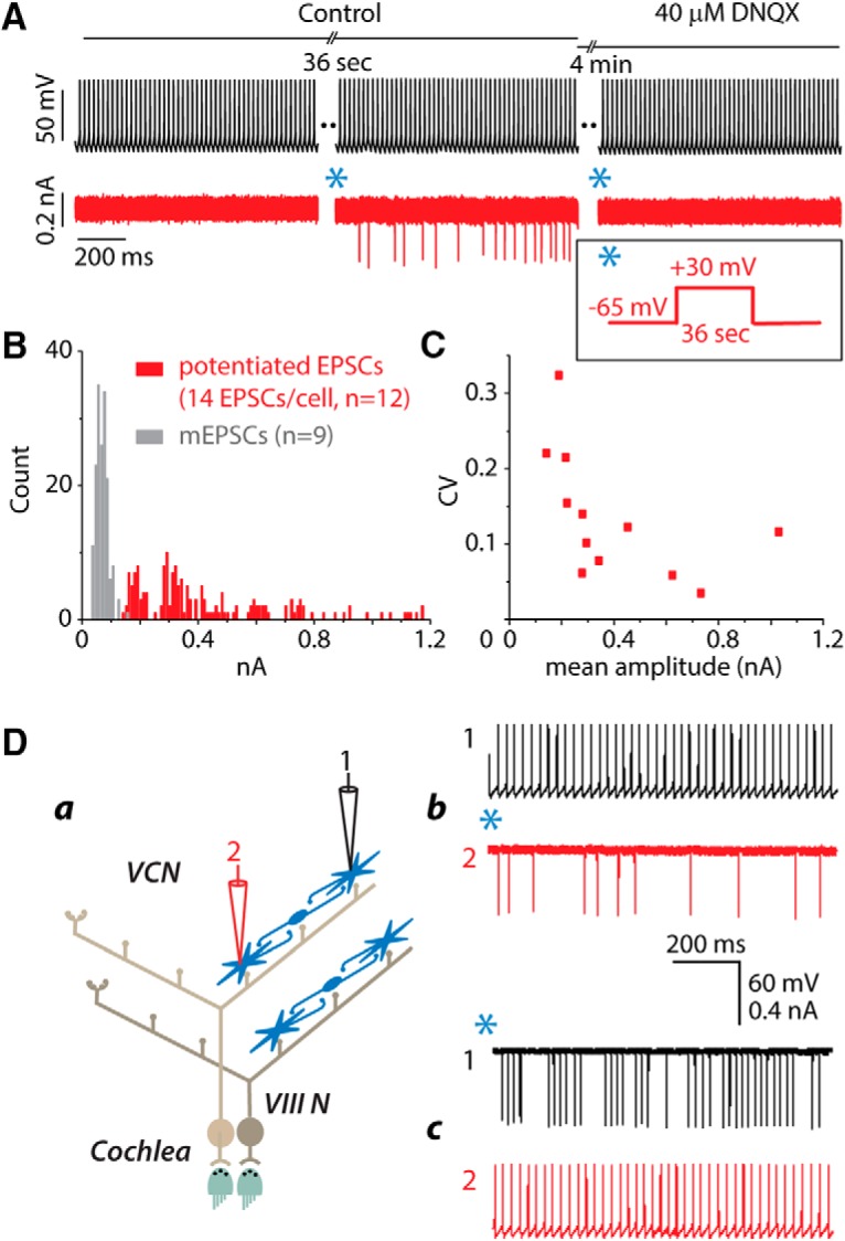
Interconnections are glutamatergic, variable in size, and bidirectional. A, After EPSCs were evoked in the postsynaptic cell and faded away, the application of 40 μm DNQX after a subsequent pairing blocked the EPSCs. B, Potentiated EPSCs evoked by stimulation of other T-stellate cells (red) are larger than mEPSCs (gray) measured from some of the same cells before potentiation. The amplitude distribution of EPSCs evoked in the postsynaptic cells of paired recordings after potentiation has peaks. To give a representative picture, only the first 14 evoked EPSCs were measured from each of 12 postsynaptic cells. C, The CV (SD/mean) of the amplitude of all evoked EPSCs in those same 12 postsynaptic cells varies. Da, Schematic diagram depicts a dual recording. Db, After pairing presynaptic firing with the postsynaptic voltage protocol shown in the inset (*), action potentials in cell 1 (black) evoked EPSCs in cell 2 (red). Dc, The roles of the two recorded cells were then reversed. After pairing action potentials in cell 2 with depolarization of cell 1(*), firing in cell 2 evoked EPSCs in cell 1. In both cells EPSCs were approximately uniform in size. Inset, *, Voltage protocol in postsynaptic cell that, when paired with presynaptic firing, resulted in potentiation.
