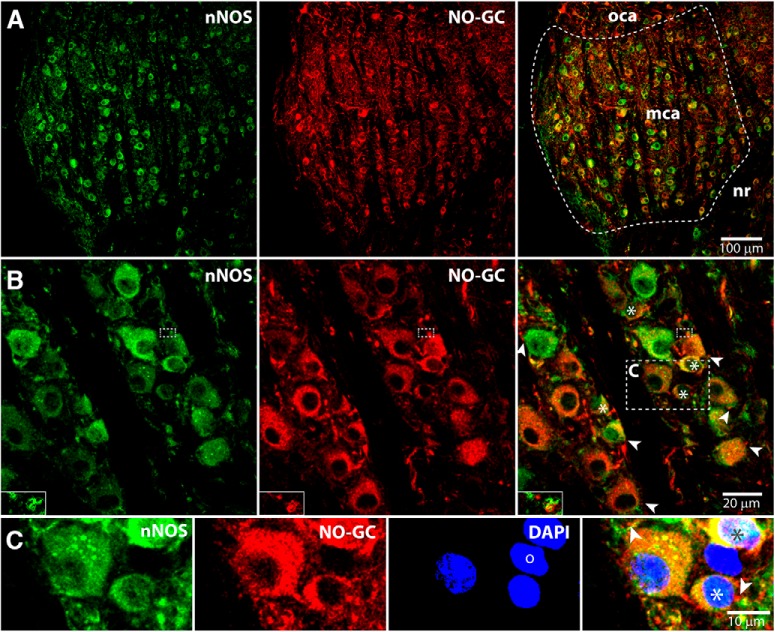
Figure 7.
Enzymes that support NO signaling are present in the pVCN where T-stellate cells reside. A, Z projection of a 5 μm stack of 0.8 μm confocal images shows a sagittal section of the multipolar cell area (mca) of the pVCN with a small part of the octopus cell area (oca) and of the nerve root (nr) also visible. In the multipolar cell area, labeled cell bodies lie between fascicles of auditory nerve fibers. nNOS (green) and NO-GC (red) are coexpressed in most cell bodies (right). B, C, Z projection of a stack (0.1 μm, ∼2.5 μm) from a section from another animal shows the labeling pattern at higher magnification. The brightness of labeling for both nNOS and NO-GC varies between cells. The right panel shows that large cell bodies, as well as small cell bodies (stars), are double labeled for nNOS and NO-GC. Puncta that are brightly labeled for NO-GC (arrowheads) can be observed apposing double-labeled large and small cell bodies. Insets in B are enlargements from single confocal images that show overlap of nNOS and NO-GC (yellow) in a punctum in the area outlined by small dashed boxes. The yellow color indicates that nNOS and NO-GC are close together. C, Enlargements of the area outlined in B showing NO-GC-labeled puncta apposed to both large and small cells. One DAPI-labeled (blue) neuron (o) was labeled for neither nNOS nor NO-GC.

