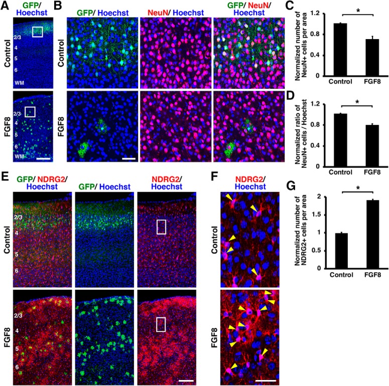
Figure 5.
Activation of FGF signaling decreased neurons and increased astrocytes in vivo. pCAG-EGFP plus either pCAG-FGF8 or pCAG control vector was introduced into the mouse cerebral cortex at E15.5 by IUE, and coronal sections were prepared at P15. Sections were stained for GFP plus either NeuN (A–D) or NDRG2 (E–G), and Hoechst 33342. A–D, FGF8 overexpression reduced layer 2/3 neurons. The areas within the boxes in A were magnified and shown in B. Fewer NeuN-positive neurons were observed in layer 2/3 of the FGF8-transfected cortex. C, Quantification of the number of NeuN-positive cells in layer 2/3. The number of layer 2/3 neurons was significantly suppressed by FGF8 (unpaired Student's t test, *p = 0.0069). D, The percentage of cells in layer 2/3 coexpressing NeuN. The percentage was significantly reduced by FGF8 (unpaired Student's t test, *p = 0.0023). E–G, FGF8 overexpression increased astrocytes. The areas within the boxes in E were magnified and shown in F. NDRG2-positive astrocytes were markedly increased by FGF8 (arrowheads). G, Quantification of the number of NDRG2-positive cells in the cerebral cortex. Astrocytes were significantly increased by FGF8 (unpaired Student's t test, *p < 0.0001). Error bars represent mean ± SEM. Scale bars: A, 200 μm; B, 50 μm; E, 100 μm; F, 20 μm. Numbers indicate layers in the cortex. WM, White matter.

