Figure 3.
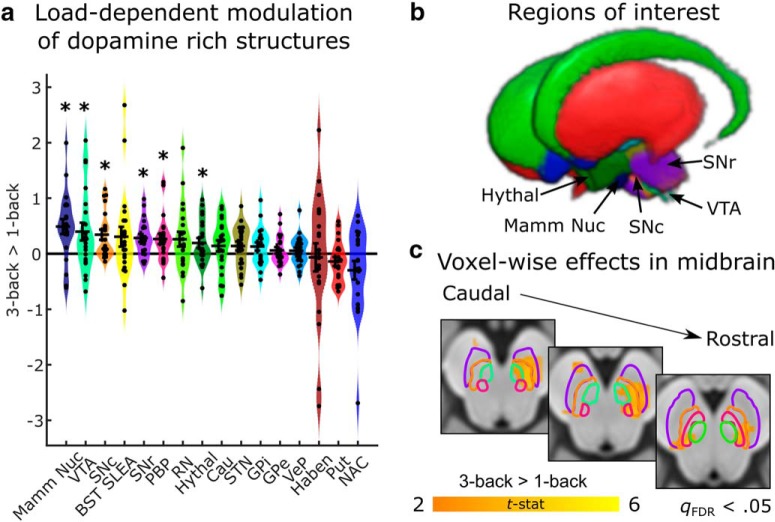
Load-dependent activity in dopamine-rich areas during a working memory task. a, ROI analysis reveals multiple subcortical and brainstem regions that are more active as working memory load increases in the N-back task. Black crosses reflect mean and SE of contrasts between the three-back and one-back conditions. Colored distributions reflect smoothed kernel density estimates of the data distribution. Individual circles indicate the value of the contrast for each subject. Asterisks denote regions that are significant at p < 0.05 uncorrected. b, Three-dimensional rendering of dopamine-rich areas (and neighboring landmark structures, i.e., extended amygdala, mammillary nucleus, and red nucleus) included in the analysis (Pauli et al., 2018). Areas that exhibited significant load-dependent effects are labeled. The parabrachial pigmented nucleus is not visible, as it is obscured by the SN (c). Mass-univariate analysis within the dopaminergic ROIs reveals multiple regions that show load-dependent effects. Color map reflects t-statistics from a one-sample t test conducted on contrasts of three-back versus one-back conditions. Orange colors indicate greater activation during the three-back task. Effects are thresholded at a voxelwise cutoff of qFDR < 0.05.

