
Figure 10.
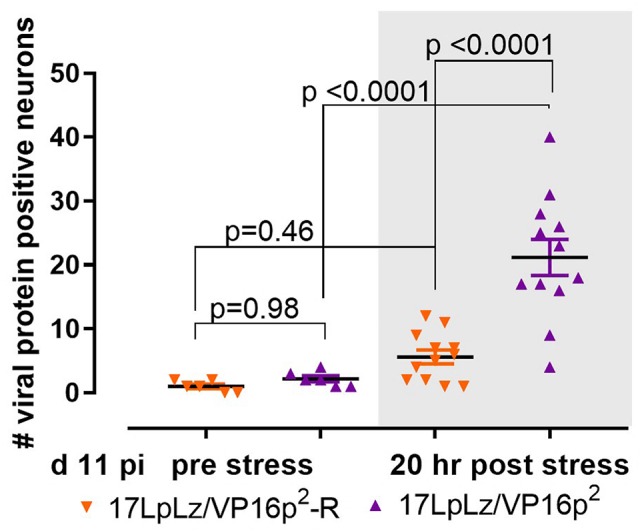
Stress induced transition into the lytic cycle. Mice were infected on scarified corneas with 1 × 10⁵ pfu of either 17LpLz/VP16p2 or its rescue17LpLzVP16p2-R. At 11 day pi, mice from each infection group were untreated or stressed (see Section “Materials and Methods”). At 20 h post stress, TG from both unstressed and stressed groups from each infection group were dissected and processed for the detection of viral proteins as detailed in Section “Materials and Methods.” The number of neurons expressing viral proteins (evidence of entry into the lytic program) in each TG was counted. Data were analyzed using ordinary one-sided ANOVA with Tukey’s multiple comparison test.