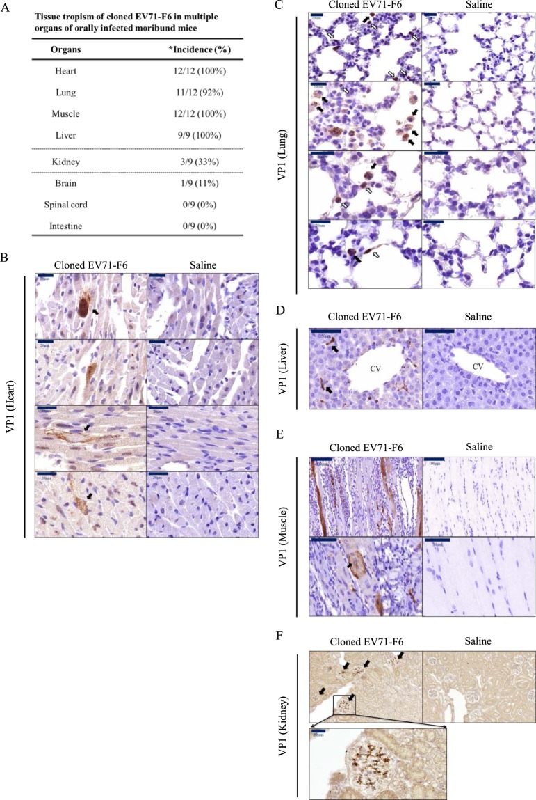
Figure 2.
Tissue distribution of EV71 in orally infected NOD/SCID mice. Experimental design was as described in Fig. 1B. Paraffin-embedded sections of different tissues from moribund mice were visualized with immunohistochemistry using a rabbit antibody specific for EV71 protein VP1. (A) A summary of EV71 tissue tropism in this oral infection model. * incidence rates represent the numbers of VP1-positive organs divided by the total number of examined moribund mice. Strong signals of VP1 can be detected in (B) cardiomyocytes, (C) alveoli (black arrow) and epithelial-like cells (white arrow) in the lung, (D) morphologically characteristic Kupffer cells around the central vein, (E) myocytes of muscle. Centralized nuclei (black arrow) is known to be associated with muscle regeneration, (F) kidney glomerulus.

