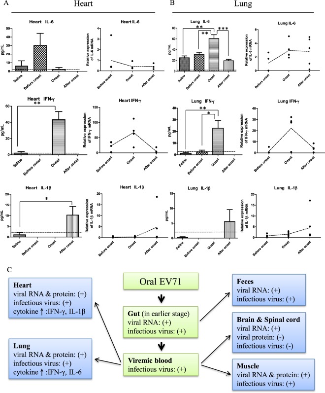
Figure 6.
Dissection of virus-host interactions in different tissues in the NOD/SCID mice infected with cloned EV71-F6. (A,B) A time course study on the expression profiles of inflammatory cytokines in the cardiopulmonary system by ELISA and RT-qPCR. Experimental design is as outlined in Fig. 1B. Sequential expression of IL6, IFN-γ, and IL-1β were detected in both heart and lung before, during, and after disease onset, respectively. In the ELISA data, dashed lines represent the cutoff value of minimal detectable dose. In the RT-qPCR data, dashed lines represent RNA expression levels relative to the saline control from an averaged value of at least 4 mice, with duplicated samples from each mouse. (C) A hypothesis of viral spreading from the oral-gut axis to peripheral tissues via blood circulation at earlier time points post-infection (data from Figs 1, 2, 6). Note: At the moribund stage, no virus was detected in the intestine (Fig. 2A).

