Fig. 6.
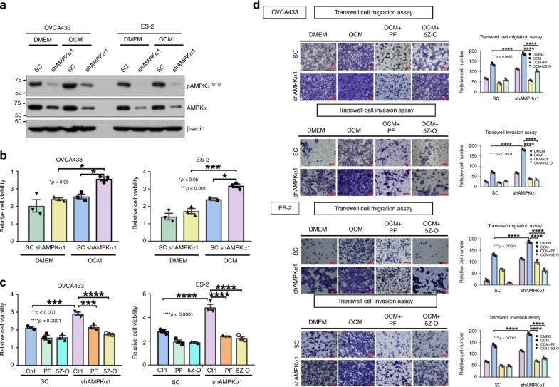
Targeting AMPK or inhibiting TAK1 signaling activity impairs ovarian cancer aggressiveness. a AMPKα1 knockdown stable clones were established via shRNA lentiviral approach targeting the α1-isoform of AMPK in OVCA433 and ES-2 cells, and cells cultured in DMEM or OCM (2 h) could not induce AMPK activity (pAMPKThr172) in shAMPKα1 clones compared with their scrambled controls (SC) of both cell lines. b XTT cell proliferation assay shows that OCM can promote proliferation of ES-2 and OVCA433 cells, but after depletion of AMPKα1 in both cell lines, shAMPKα1 clones show significantly higher cell proliferation than that of scrambled controls (SC) cultured in DMEM or OCM for 3 days. c XTT cell proliferation assay shows that 3 days of co-treatment with AMPK activator PF-06409577 (50 μM) (PF) or TAK1 inhibitor (5Z)-7-Oxozeaenol (2.5 μM) (5Z-O) significantly inhibited cell proliferation in both parental and AMPKα knockdown clones compared with the scrambled controls (SC) of ES-2 and OVCA433 cells. d Transwell cell migration/invasion assays demonstrate that the cell migratory and invasive capacities of OVCA433 and ES-2 cells are enhanced by OCM. Knockdown of AMPKα1 further promotes the migration and invasion rates of cells cultured in OCM or DMEM. In contrast, co-treatment with AMPK activator PF-06409577 (50 μM) (PF) or TAK1 inhibitor 5Z-O (2.5 μM) somewhat abrogated the cell migratory and invasive capacities of AMPKα1 knockdown clones and scrambled controls (SC) of both cell lines. The stained cells were counted at least from four randomly selected fields. Representative images and quantitative results were shown by bar charts. Scale bar = 50 μm

