Fig. 7.
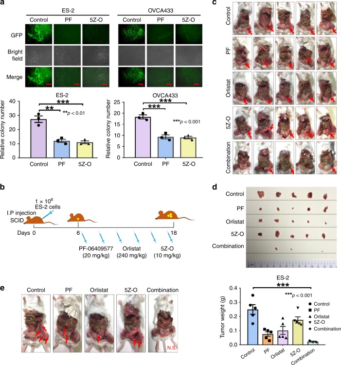
AMPK/FASN/TAK1/NF-κB signaling axis is required for metastatic colonization. a eGFP-labeled ES-2 and OVCA433 ovarian cancer cells were established by infection with LV-CMV-RLuc-IRES-GFP lentiviral particles. After incubating GFP-labeled ovarian cancer cells with omental tissues from 6- to 8-week-old SCID female mice, the results show that ES-2 and OVCA433 cells exhibit a significant number of tumor colonies on murine omenta on day 30. However, co-treatment with AMPK activator PF-06409577 (50 μM) (PF) or TAK1 inhibitor 5Z-O (2.5 μM), remarkably reduces the number and size of tumor colonies by 45–55% on the murine omenta. Scale bar = 100 μm. b Schematic overview showing the experimental protocol of the anti-tumorigenic effect of the combined cocktail of the AMPK activator PF-06409577 (20 mg/kg), FASN inhibitor orlistat (240 mg/kg), and TAK1 inhibitor 5Z-O (10 mg/kg) on ovarian cancer cells in SCID mice. ES-2 cells (1 × 10⁶/200 µl) were injected into the intraperitoneal cavity of 5–6-week-old SCID female mice. On day 6, the above three drug reagents were injected individually or in combination (for a total of six injections) from day 6 to day 18. c Images show tumor formation in all mice. d Images showing tumor nodules obtained from all mice. The bar chart shows the average tumor weight obtained from each group. e Representative images showing the number and locations of tumor nodules distributed in the intraperitoneal cavity of each mouse group. Results were presented as mean ± S.E.M. Data were analyzed by one-way ANOVA with Tukey’s post hoc test (*p < 0.05, **p < 0.01, ***p < 0.001, ****p < 0.0001)

