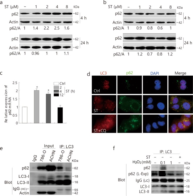
Figure 3.
ST increases the expression of p62 in 786-O cells. 786-O (a) and ACHN (b) cells were treated with ST (0–8 μM/L) for up to 24 h. Cells were lysed and subjected to immunoblotting (8–13% PAGE) with the indicated antibodies. Actin was used as a loading control. (c) qPCR was performed to detect the mRNA expression of p62 following ST treatment for up to 24 h in 786-O cells. (d) 786-O cells were split onto coverslips, cultured overnight, and treated with ST (4 μM/L) or together with CQ (15 μM/L). They were then fixed with 4% paraformaldehyde, and images were obtained by fluorescence microscopy after labeling with anti-LC3 and p62 antibodies and staining with DAPI. (e) IgG2b (rabbit) was used as a negative control. ACHN and 786-O cells were lysed and precipitated using the antibody against LC3. (f) Following treatment with ST (8 μM/L: 4 h) or H2O2 (0.1, 1 mM/L: 2 h) for up to 4 h, 786-O cells were lysed and precipitated using the antibody against LC3. The immunoprecipitates were resolved by electrophoresis and probed by immunoblotting (8–13% PAGE) with the indicated antibodies. The results were similar among experiments repeated at least twice. (L-Exp: long exposure; HC: heavy chain; LC: light chain; Actin: A; Input: whole cell lysate).

