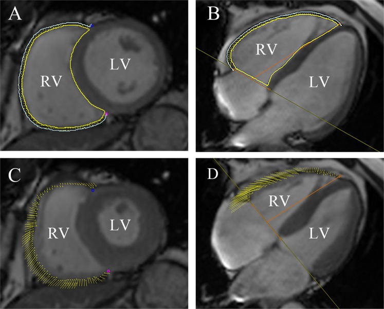
Figure 1.
CMR feature tracking using cmr42 (Circle Cardiovascular Imaging Inc., Calgary, Canada) in short-axis, and four-chamber long-axis cine images at the end-diastole (A,B) and end-systole (C,D). The yellow and cyan curves delineate the endocardial and epicardial contours, respectively. The yellow dots represent the right ventricle myocardial voxel points, and the yellow short line on the images shows the tracking of the ventricle myocardial voxel points. The orange line is used to define the base and apex of tricuspid valve plane, and the apical plane. Abbreviations: CMR, cardiovascular magnetic resonance; T2DM: type 2 diabetes mellitus.

