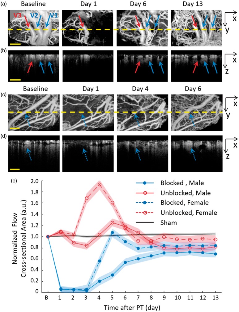
Figure 5.
Dynamic blood flow observation of dMCA after PT occlusion. Male: (a) Two of the dMCAs were completely blocked after PT (indicated with blue arrows) in the irradiation core and one vessel in the peripheral area (the red arrow) was less infracted. (b) The cross sections of blood flow imaging of yellow dot line in (a). The recovery blood flow of dMCA (blue arrows) after PT was in the original position and depth as Baseline. Female: (c) The dMCAs was completely blocked after PT (indicated with the blue dot arrows) in the irradiation core. (d) The cross sections of blood flow imaging of yellow dot line in (c). (e) Average flow cross-sectional area (FCA) changes of blocked dMCA in the core ischemia region and the unblocked in the peripheral area of the male and female rats. dMCA: distal middle cerebral arteries.

