Figure 4.
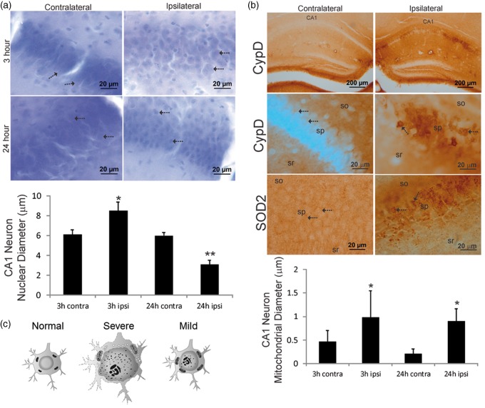
Hippocampal CA1 neuron nuclear (a) and mitochondrial (b) pathology occurred early after HI in neonatal mouse brains with mild edema. (a) Nissl staining of CA1 at 3 h after HI showed that the nuclei (arrows) of ipsilateral CA1 neurons were swollen compared contralateral side and there was ipsilateral parenchymal edema. At 24 h, the nuclei (arrows) of ipsilateral CA1 neurons were shrunken compared contralateral CA1 neurons, while parenchymal edema persisted. The graph at the bottom illustrates CA1 neuron nuclear diameters at 3 h (n = 5) and 24 h (n = 6) after HI (mean ± SD, *p < 0.001; **p < 0.0001). (b) Top row: low magnification images showing that CypD immunoreactivity selectively accumulated in the ipsilateral CA1 pyramidal neuron cell layer at 3 h after HI. In the contralateral CA1, there was diffuse neuropil CypD immunoreactivity. Second row: ipsilateral CA1 pyramidal neuronal cell bodies markedly accumulated large CypD positive mitochondria (arrows), in contrast to the very fine particles of CypD staining (arrows) in the contralateral neurons. sr: stratum radiatum, sp: stratum pyramidal, so: stratum oriens. Third row: SOD2 immunoreactivity showed a different pattern of mitochondrial staining that was more generally localized to mitochondria throughout the sp (arrows), so, and sr. In the ipsilateral CA1 neurons, a subset of mitochondria showed swelling (arrows). The graph at the bottom illustrates CA1 neuron cell body mitochondrial diameters at 3 h (n = 5) and 24 h (n = 6) after HI in mice with mild edema (3 h, *p < 0.05; 24 h *p < 0.001). (c) A diagram illustrating representative cellular and sub-cellular structural changes in the hippocampal neurons at 3 h after HI.

