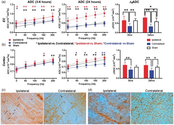
Figure 5.
dMRI measurements of external capsule (EC) and cortex of mouse brains with mild edema at 3–6 and 24 h post-injury and MAP2 immunostaining that revealed early neocortical structural pathology at 3 h after HI. (a–b) ADC and ΔfADC measurements in the EC and cortex in mild edema cases at the 3–6 and 24-h time points. PG-dMRI measurements are denoted as 0 Hz. * and ** denote significant differences with p-values less than 0.05 and 0.005, respectively. Black, red, and blue asterisks denote the statistical differences between ipsilateral and contralateral ROIs of the HI-injured mice, between ipsilateral and sham groups, and between contralateral and sham groups, respectively, using pairwise t-tests with Bonferroni correction following two-way ANOVA analysis. Numerical values of PGSE-ADC and ΔfADC can be found in Tables 1 and 2. (c) MAP2-stained images of ipsilateral (injured) and contralateral (non-HI) parasagittal parietal cortex showing large MAP2-positive dendritic bundles of radially oriented neocortical dendrites. In the ipsilateral cortex, the dendrites were bundled (brackets) and some dendrites appeared tortuous and swollen (arrows). The neuropil was interspersed with pale zones (asterisks). The contralateral cortex had less apparent dendritic bundling (arrows). (d) Ipsilateral neocortical dendrites were thicker than contralateral neocortical dendrites (arrows) as indicated by their width relative to the scale bars.

