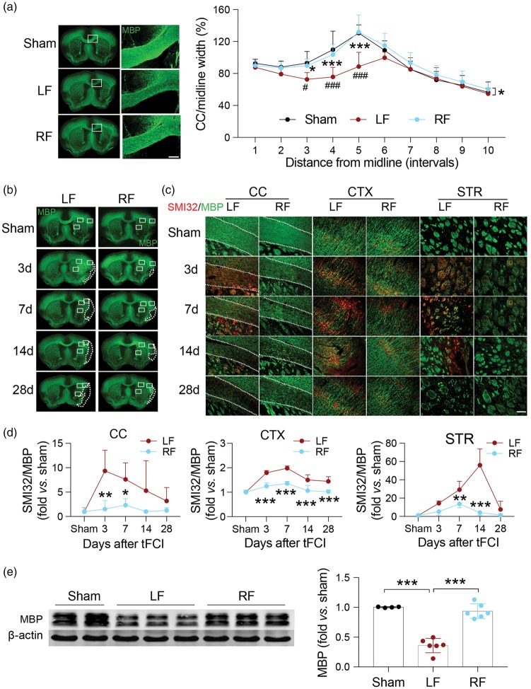
Figure 2.
White matter integrity is better preserved in ischemic mice with food restriction. (a) Representative images of MBP immunofluorescent staining of sham (top row) and tFCI mice given LF or RF (middle and bottom rows, respectively) 28 days after ischemic insult. The ischemia-induced reduction in CC thickness is prevented by RF. The white boxes indicate areas that were enlarged in high-power images (right column, scale bar = 200 µm). Quantification of the width of the CC indicates that RF maintained the width of CC 28 days after tFCI. n = 4–7 for each group. *p ≤ 0.05, ***p ≤ 0.001 vs. LF; #p ≤ 0.05, ###p ≤ 0.001 vs. sham. (b) Representative immunofluorescent images of sections from each treatment group stained with MBP. Dashed line depicts brain infarct. (c) Representative high-power images of regions depicted in white rectangles in (b) from each treatment stained with MBP (green) and SMI32 (red). Dashed line depicts the border of the CC. Scale bar = 100 µm. (d) Quantification of the ratio of SMI32/MBP fluorescence intensity in corpus callosum (CC), cortex (CTX) and striatum (STR) of the ipsilateral hemisphere after tFCI indicates that RF-feeding preserves white matter integrity in ischemic mice. n = 5 mice/group at each time point. *p ≤ 0.05, **p ≤ 0.01, ***p ≤ 0.001 vs. LF. (e) Representative image of Western blot for MBP in corpus callosum of ipsilateral hemisphere (left panel). β-Actin was used as loading control. Semi-quantification of bands relative to sham mice (right panel) shows that RF prevented ischemia-induced loss of MBP. n = 4–6/group. ***p ≤ 0.001. All data are presented as mean ± SD.

