Figure 1.
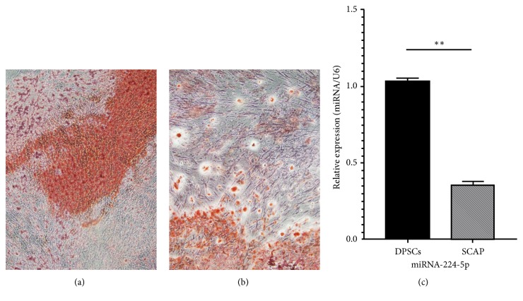
Osteogenic differentiation for 21 days. Red areas are calcified nodules. (a) Stem cells from the apical papilla (SCAP). (b) Human dental pulp stem cells (DPSCs). There are more calcified nodules in picture (a) than in picture (b). Original magnification: 40×. (c) Quantitative RT-PCR showing the levels of miR-224-5p expression in DPSCs and SCAP. Values are expressed as relative expression with respect to the endogenous control gene U6 (2−ΔΔCt). Data represent the mean±SEM of 3 independent experiments conducted in duplicate, ∗∗P<0.01.

