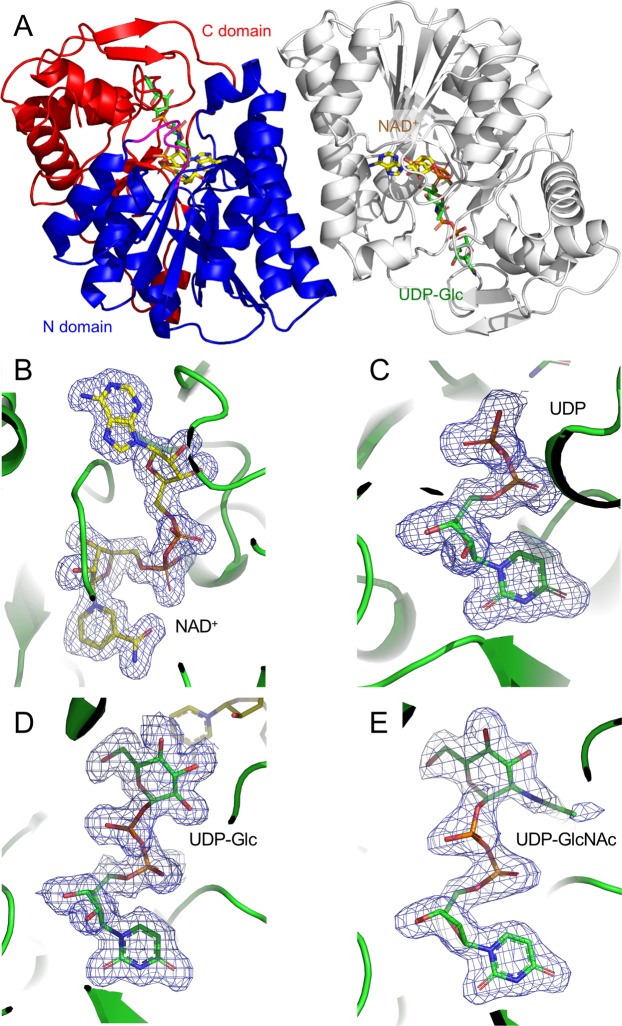
Figure 3.
Crystal structure of bGalE. (A) Overall structure of the biological dimer assembly. One protomer is shown in blue (N domain, residues 1–177 and 236–262) and red (C domain, residues 178–235 and 263–340), and the other symmetry-related protomer is shown in gray. mFo-DFc omit map of NAD+ (B) and UDP (C) in the NAD+ + UDP complex, UDP-Glc (D), and UDP-GlcNAc (E) are shown with a contour level of 3.0σ. NAD+ and UDP or UDP-sugar molecules are shown with yellow and green sticks, respectively.

