


Figure 2.
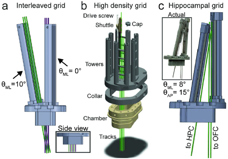
a) Linear dense interleaved grid in coronal profile; inset shows side view. b) High density grid. One shuttle, screw, and cap shown for clarity. Green tracks show electrode trajectories. Grid footprint slots into mating collar that is mounted onto chamber. c) Hippocampal grid render; inset shows photo of assembled system.