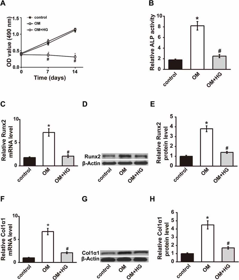
FIGURE 1.

T2DM model was established in MC3T3-E1 cells. (A) Cell viability was measured by CCK-8 assay 0, 7 and 14 days after combined treatments with OM and HG. ALP activity (B), Runx2 expression (C-E) and Col1α1 expression (F-H) were measured 14 days after OM and HG treatment. OM, osteogenic medium; HG, high glucose-free fatty acids; ALP, alkaline phosphatase; Runx2, runt-related transcription factor 2; Col1α1, collagen type I α 1. Control, MC3T3-E1 cells cultured in α-MEM with 10% FBS for 14 days. * P < .01, compared with the control group (growth medium); # P < .01, compared with OM group.
