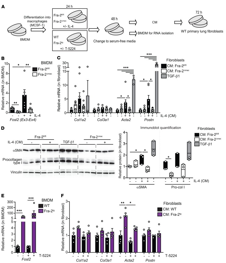
Figure 5. CM from Fra-2–expressing BM-derived macrophages induces lung fibroblast activation.
(A) Experimental design to assess the effect of BMDM conditioned medium on WT primary lung fibroblasts. Experiment was repeated 5 and 2 times for the Fra-2 loss and gain of function, respectively. Each individual value represents a biological replicate, since each BMDM culture originates from 1 individual mouse. IL-4 was added at 20 ng/mL, T-5224 at 3 μM, and TGF-β1 at 0.5 ng/mL. (B) Fra-2 expression in BMDMs when CM was collected (qRT-PCR). Note that specific primers located in the floxed/deleted exons (Ex3-Ex4) are used. *P < 0.05; **P < 0.01, 1-way ANOVA; Bonferroni’s post test. (C) qRT-PCR analysis of fibroblast marker genes in primary WT lung fibroblasts cultured with CM and TGF-β1 (positive control). *P < 0.05; ***P < 0.001, 1-way ANOVA; Bonferroni’s post test. Group analysis for each gene. TGF-β1, n = 3; other groups, n ≥ 7. (D) Immunoblot analysis of procollagen I and α-SMA in primary lung fibroblast lysates. Relative densitometry quantification for each protein is shown as a ratio to vinculin density (loading control). Individual values and mean ± SEM from 1 experiment are plotted. *P < 0.05, unpaired t test; 1-tailed. Pro-col I, procollagen I. (E) Fra-2 expression in WT and Fra-2Tg BMDMs at the time the CM was collected (qRT-PCR). ***P < 0.001, 1-way ANOVA; Bonferroni’s post test. (F) qRT-PCR analysis of fibroblast marker genes in primary WT lung fibroblasts cultured with WT and Fra-2Tg BMDM-CM. *P < 0.05; **P < 0.01, paired 2-tailed t test. In all panels, bars represent mean ± SD/SEM. Relative mRNA and protein expression in untreated Fra-2fl/fl BMDMs and derived CM is set to 1.

