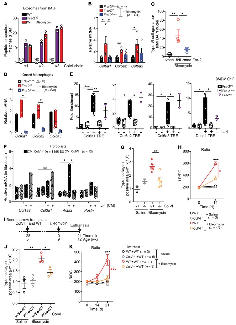
Figure 6. ColVI expression contributes to lung fibrosis.
(A) Peptide-spectrum match (PSM) identified for ColVI fragments by LC-MS/MS of BALF exosomes extracted from 12-week-old WT mice, Fra-2Tg mice, and WT mice after 12 days of bleomycin treatment. ND, not detected. (B) ColVI gene expression in lungs 21 days after treatment. Relative expression in saline-treated Fra-2fl/fl is set to 1. *P < 0.05; **P < 0.01, unpaired 1-tailed t test. (C) ColVI-positive area in lungs 21 days after treatment. Control group was treated with saline. Data were normalized to nuclei number. *P < 0.05; **P < 0.01, 1-way ANOVA; Bonferroni’s post test. (D) ColVI gene expression in sorted nonalveolar lung macrophages 14 days after bleomycin treatment. Expression in sorted cells from saline-treated lungs is set to 1. *P < 0.05, unpaired 2-tailed t test. (E) Fra-2 ChIP assay in BMDM. Experiment was repeated twice. Relative expression in untreated Fra-2fl/fl cells is set to 1. *P < 0.05; **P < 0.01; ***P < 0.001, unpaired 1-tailed t test. (F) Fibroblast marker gene expression in WT lung fibroblasts (CM, BMDM CM). Experiment was repeated twice. *P < 0.05; **P < 0.01, 1-way ANOVA; Bonferroni’s post test. (G) Type I collagen area of lungs 14 days after saline or bleomycin treatment. Data from 2 independent experiments are plotted. **P < 0.01, unpaired 2-tailed t test. (H) Respiratory function 14 days after saline or bleomycin treatment. Two independent sets of mice were treated. *P < 0.05; ***P < 0.001, 2-way ANOVA; Bonferroni’s post test. (I) Schematic for experimental design and time line of saline- and bleomycin-treated WT mice transplanted with either WT BM (WT→WT) or ColVI–/– BM (KO→WT). Bleomycin was injected into 2 independent sets of mice. (J) Type I collagen area at the end of the experiment. *P < 0.05; **P < 0.01, unpaired 1-tailed t test. (K) Respiratory function 21 days after bleomycin. ***P < 0.001, 2-way ANOVA; Bonferroni’s post test.

