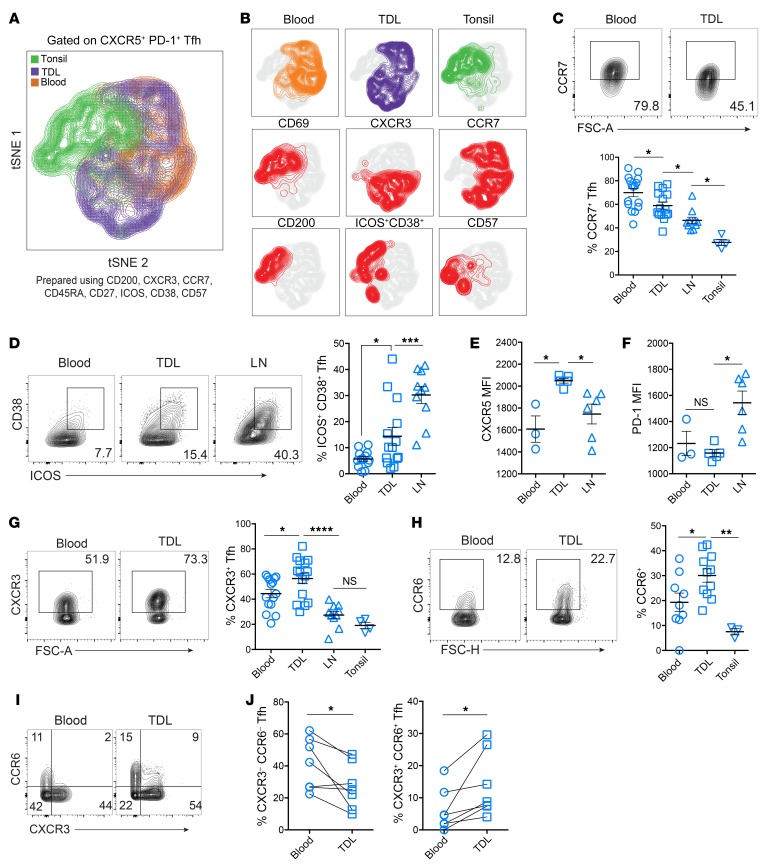
Figure 3. TDL Tfh have an intermediate activation phenotype between LNs and blood, with increased expression of chemokine receptors.
(A) tSNE analysis of Tfh (all CXCR5+PD-1+) from tonsils (n = 4), TDL (n = 5), and blood (n = 5) using the indicated 8 surface markers. (B) Plots of individual marker expression within the tSNE space. (C) Frequency of CCR7+ Tfh in blood (n = 16), TDL (n = 15), LNs (n = 10), and tonsil (n = 4). (D) Frequency of ICOS+CD38+ Tfh in blood (n = 13), TDL (n = 14), and LNs (n = 10) (E and F) CXCR5 and PD-1 MFI of Tfh in blood (n = 3), TDL (n = 5), and LNs (n = 7). (G) Frequency of CXCR3 in blood (n = 15), TDL (n = 15), LNs (n = 10), and tonsil (n = 4). (H) CCR6 expression in paired blood and TDL (n = 7). (I) Expression of CXCR3 versus CCR6 by compartment in paired samples (n = 7), with (J) frequencies of Tfh expressing neither or both chemokine receptors. Error is reported as SD. Comparisons shown in C, D, G, and H were performed using ANOVA with the Holm-Šídák post-test. Comparisons shown in E and F were performed using Kruskal-Wallace with Dunn’s post-test. Paired 2-tailed t tests were performed for J. *P < 0.05; **P < 0.01; ***P < 0.001; ****P < 0.0001.

