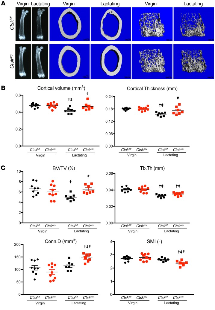
Figure 3. Deletion of Ctsk in osteocytes preserves cortical and cancellous bone during lactation.
(A) Representative x-ray and μCT images of virgin and lactating Ctskfl/fl and Ctskocy femurs (n = 7–9 per group). Scale bars: 100 μm. (B) μCT analysis of cortical bone at the femoral midshaft of virgin and lactating Ctskfl/fl mice (black dots and squares, respectively) and Ctskocy mice (red dots and squares, respectively) (n = 7–9 per group). (C) μCT analysis of trabecular bone at the distal femur of virgin and lactating Ctskfl/fl (black dots and squares, respectively) and Ctskocy (red dots and squares, respectively) mice (n = 7–9 per group). Results represent the mean ± SEM. †P < 0.05 versus virgin Ctskfl/fl mice; ‡P < 0.05 versus virgin Ctskocy mice; and #P < 0.05 versus lactating Ctskfl/fl mice; 2-way ANOVA followed by Fisher’s PLSD test.

