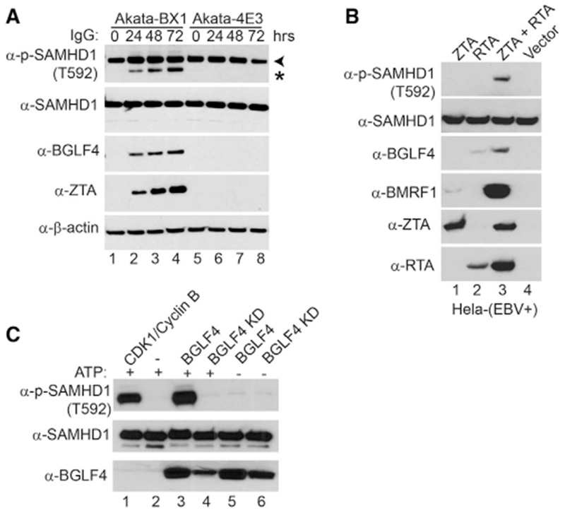
Figure 2. SAMHD1 Is Phosphorylated by EBV Protein Kinase BGLF4.

(A) SAMHD1 is phosphorylated upon lytic induction of EBV. Western blot analysis was performed on cell lysates from Akata-BX1 (EBV+) and Akata 4E3 (EBV−) cells using antibodies as indicated. The cells were either untreated (0 hr) or treated with anti-human IgG for 24, 48, or 72 h to induce lytic reactivation. Arrowhead denotes the major 72-kDa phospho-SAMHD1 band, and asterisk denotes a 69-kDa non-specific band or a phospho-SAMHD1 band derived from a SAMHD1 isoform protein.
(B) SAMHD1 is phosphorylated in EBV-replicating HeLa cells. Western blot analysis was performed on cell lysates from HeLa (EBV+) transfected with EBV ZTA, RTA, or ZTA plus RTA as indicated for 48 h to induce lytic reactivation. The phosphorylation of SAMHD1 correlated with BGLF4 expression.
(C) SAMHD1 is phosphorylated by EBV protein kinase BGLF4 in vitro. Recombinant SAMHD1 protein was mixed with purified wild-type or KD BGLF4 for 30 min at 30°C. As a positive control, SAMHD1 was incubated with CDK1/Cyclin B (lane 1), which is a known kinase for SAMHD1. As negative controls, either kinase or ATP was omitted in the reaction mixture (lanes 2, 5, and 6). The phospo-SAMHD1, SAMHD1, and BGLF4 were detected by western blot using antibodies as indicated.
See also Figure S1.
