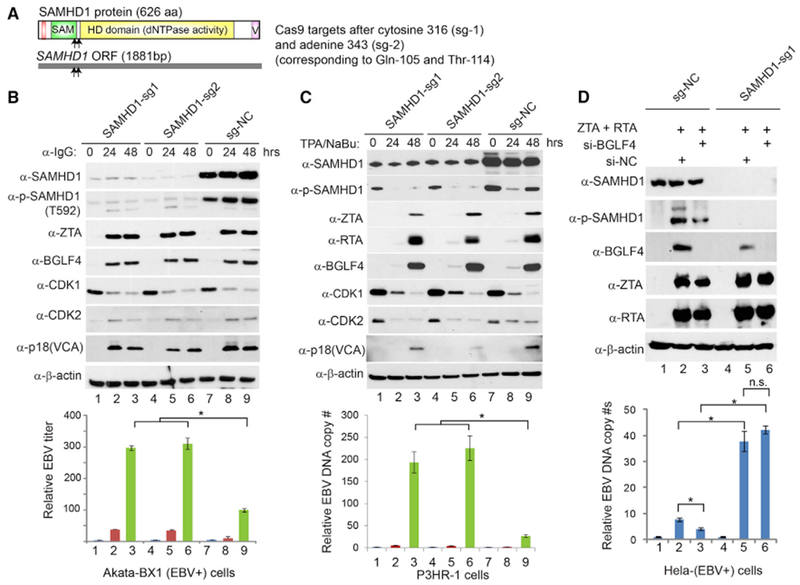
Figure 3. SAMHD1 Depletion Facilitates EBV Lytic Replication.

(A) Schematic representation of Cas9 target sites within the 1,881-bp SAMHD1 open read frame (ORF). The corresponding positions of amino acids are also shown.
(B) SAMHD1 depletion facilitates EBV replication in Akata cells. SAMHD1-depleted (sg-1 and sg-2) and control (sg-NC) Akata-BX1 (EBV+) B cells were treated with IgG cross-linking to induce EBV lytic reactivation. Western blot analysis was performed using antibodies as indicated. β-Actin served as a loading control. Relative EBV titer of lytically induced Akata-BX1 (EBV+) cells carrying SAMHD1-deletion or control was measured using a Raji cell infection assay.
(C) SAMHD1 depletion facilitates EBV replication in P3HR-1 cells. SAMHD1-depleted (sg-1 and sg-2) and control (sg-NC) P3HR-1 cells were treated with TPA and sodium butyrate (NaBu) to induce EBV lytic reactivation. Western blots were performed using antibodies as indicated. β-Actin served as a loading control. Relative EBV DNA copy numbers were determined by qPCR using primers to BALF5 gene normalized by β-actin.
(D) BGLF4 knockdown suppresses EBV replication in SAMHD1-expressing cells, but not in SAMHD1-depleted cells. Control (sg-NC) and SAMHD1-depleted (sg-1) HeLa (EBV+) cells were transfected with ZTA plus RTA to induce EBV lytic reactivation. The siBGLF4-expressing plasmid was co-transfected to knock down BGLF4. The transfection of si-NC served as a negative control for siBGLF4. The EBV genome copy numbers were measured by qPCR using primers specific to EBV BALF5 gene normalized by β-actin.
Representative results from three biological replicates are presented. Data are represented as mean ± SD of technical replicates (n = 3). *p < 0.05. n.s., not significant. See also Figures S1–S4.
