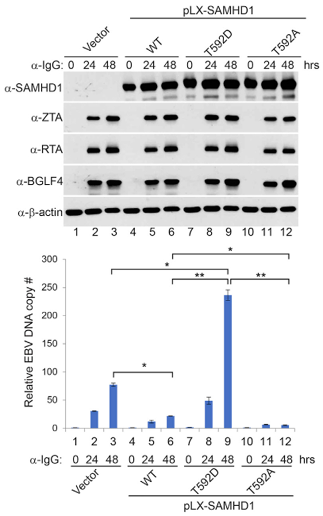
Figure 4. SAMHD1 Reconstitution Suppresses EBV Lytic Replication.

SAMHD1-depleted (sg1) Akata (EBV+) cells were used to establish SAMHD1-expressing stable cell lines using pLX-304 lentiviral constructs containing wild-type (WT), T592D, and T592A SAMHD1. Western blot analyses show SAMHD1, ZTA, RTA, and BGLF4 expression levels in different cell lines upon IgG cross-linking as indicated. Viral DNA replication was measured by qPCR using primersto BALF5. Representative results from three biological replicates are presented. The value of Vector control at 0 h (lane 1) was set as 1. Data are represented as mean ± SD of technical replicates (n = 3). *p < 0.05; **p < 0.001. See also Figure S4.
