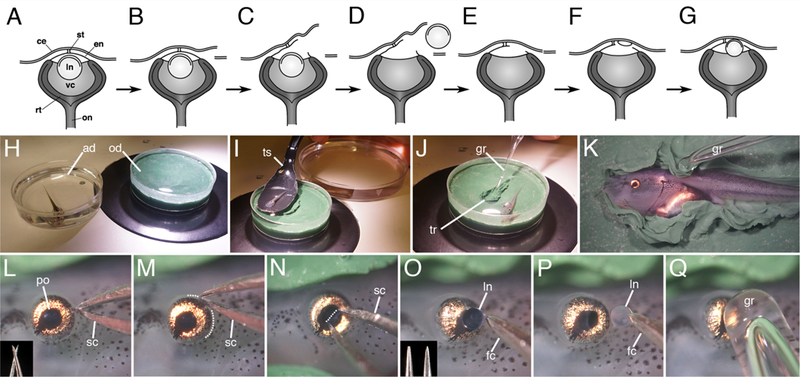
Figure 1.

Lentectomy in Xenopus laevis larvae. (A-G) Simple lentectomy diagram. H) Tadpole is anesthetized in a dish containing MS222 solution, transferred to the operating dish (I) following sedation, and immobilized on its side in a clay trough (J). Flaps of clay are used to secure the tadpole (K), and lentectomy begins with an incision on the posterior side of the eye (L-M; microscissors are shown in the inset, dotted line outlines the incision). N) An incision is made across the pupillary opening in the cornea endothelium and the intact lens capsule containing the lens cells is removed (O-P). Inset in O shows forceps prior to (left) and after sharpening (right). (Q). The cornea epithelium is smoothed over the eye with a glass rod tool. ad, anesthetic dish; ce, cornea epithelium; e, eye; en, cornea endothelium; fc, forceps; gr, glass rod; ln, lens; od, operating dish; on, optic nerve; po, pupillary opening; rt, retina; sc, scissors; st, connecting stalk; ts, transfer spoon; tr, trough; vc, vitreous chamber.
