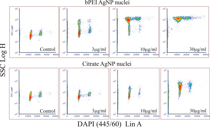
Fig 5. Flow cytometry cytograms of nuclei treated with AgNP-bPEI and AgNP-CIT.
Flow cytometry comparison of DAPI stained nuclei derived from cells treated with AgNP-bPEI or AgNP-CIT at doses between 1μg/ml and 30 μg/ml. Each successive higher dose resulted in greater amount of side scatter. This was presumably due to higher uptake of AgNP as shown in the cell scatter data (Figs 1 and 2) and the lack of complete cell lysis at all doses.

