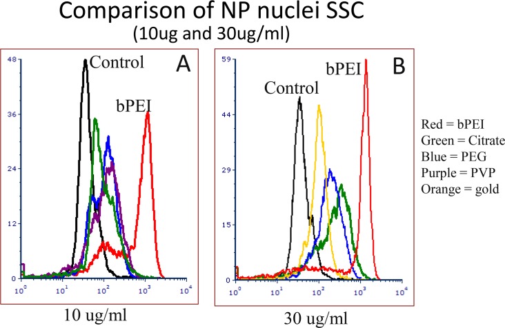
Fig 6. Comparison of nuclei SSC histograms from NP treated cells.
Histograms of nuclei derived from cells treated with the 4 types of AgNP at 10 μg/ml (Fig 6A) and 30 μg/ml (Fig 6B). All samples demonstrated an increase of scatter after AgNP incubation for 24 hours at doses between 1 and 30 μg/ml. All the AgNP showed greater SSC than the AuNP.

