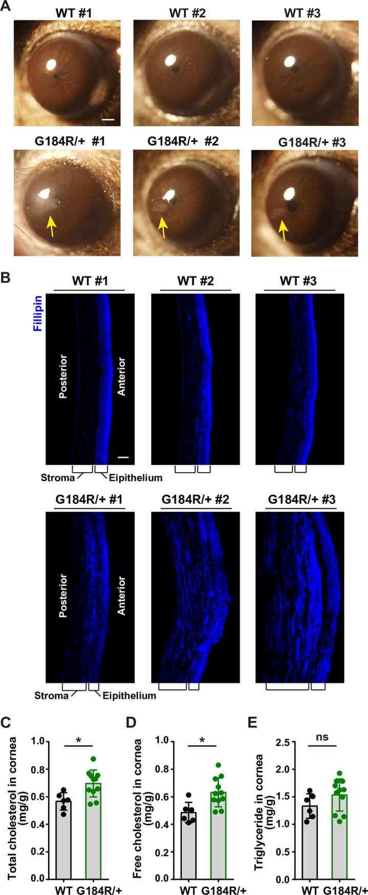
Fig 7. Accumulation of free cholesterol in the cornea of Ubiad1G184R/+ aged mice.

(A) Aged Ubiad1G184R/+ mice show corneal opacification. Male and female 102-week to 108-week old (average: 105-week) WT (17 mice/group) and Ubiad1G184R/+ (33 mice/group) littermates were fed ad libitum chow diet. Mice were first anaesthetized with pentobarbital sodium, then corneal opacifications were observed and images were captured with Olympus stereomicroscope. Yellow arrow indicates the corneal opacification. Scale bar, 200 μm.(B) Filipin staining of corneas from WT and Ubiad1G184R/+ mice. Whole eyes from aged mice in (A) were dissected and sectioned at 7 μm per slide using frozen section method. Slides were stained with Filipin to visualize free cholesterol. Scale bar, 20 μm. (C-E) Male aged WT (6 mice/group) and Ubiad1G184R/+ (11 mice/group) mice from (A) were sacrificed and excised the cornea under stereomicroscope. The lipids in corneas were extracted and measured with corresponding colorimetric kits. Values are means ± SD; p value was calculated with Student’s t test; ns, no significance. The levels of total cholesterol (C), free cholesterol (D), and triglyceride (E) in corneas from WT and Ubiad1G184R/+mice. The experiments are repeated three times and representative data are shown.
