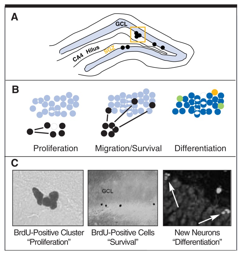
Adult neurogenesis in the dentate gyrus. (A) Neural stem cells proliferate along the subgranular zone of the dentate gyrus (shown schematically in the top panel). (B) As shown in the cartoon panels, newborn, BrdU-labeled cells (shown in black) proliferate (left), then migrate into the granule cell layer (middle), and eventually differentiate (right) into neurons (blue), glia or oligodendrocytes. As shown in the right-most cartoon panel, cell type, or differentiation, is detected by labeling for multiple proteins/markers in the same tissue. For example, we stain brain tissue sections for cell proliferation (BrdU marker, yellow) and neuron-specific proteins (blue). In the differentiation panel, green cells represent simultaneous labeling of BrdU for newborn cells (in this panel, BrdU is labeled in yellow, rather than black) and for a mature neuron marker (blue). Overlapping blue and yellow make green; these green cells represent recently formed mature neurons in adults. (C) Representative photomicrographs (bottom panel) show examples corresponding to each cartoon.

An official website of the United States government
Here's how you know
Official websites use .gov
A
.gov website belongs to an official
government organization in the United States.
Secure .gov websites use HTTPS
A lock (
) or https:// means you've safely
connected to the .gov website. Share sensitive
information only on official, secure websites.
