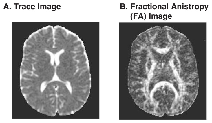
Figure 3.
Two types of diffusion tensor imaging. (A) The trace image reflects the total amount of diffusion occurring in each region and highlights the ventricles, with little difference between white and gray matter. (B) The fractional anisotropy (FA) image highlights regions where diffusion is oriented in a single direction. The ventricles and gray matter are dark, whereas the white matter tracts are bright.

