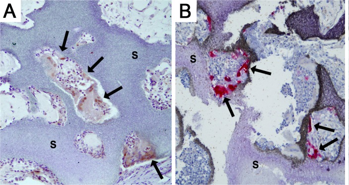
Fig 5. Characterization of the cells expressing BMP-2.
BMP-2 immunohistochemistry (A) and TRAP staining for cells of the macrophage lineage (B) were performed on sections of the same group (pBMP-2 + MSCs). Cells aligning the scaffold, indicated with the arrows, are stained BMP-2 positive (brown in A) and TRAP positive (red in B). s = scaffold. Scale bar = 200 μm.

