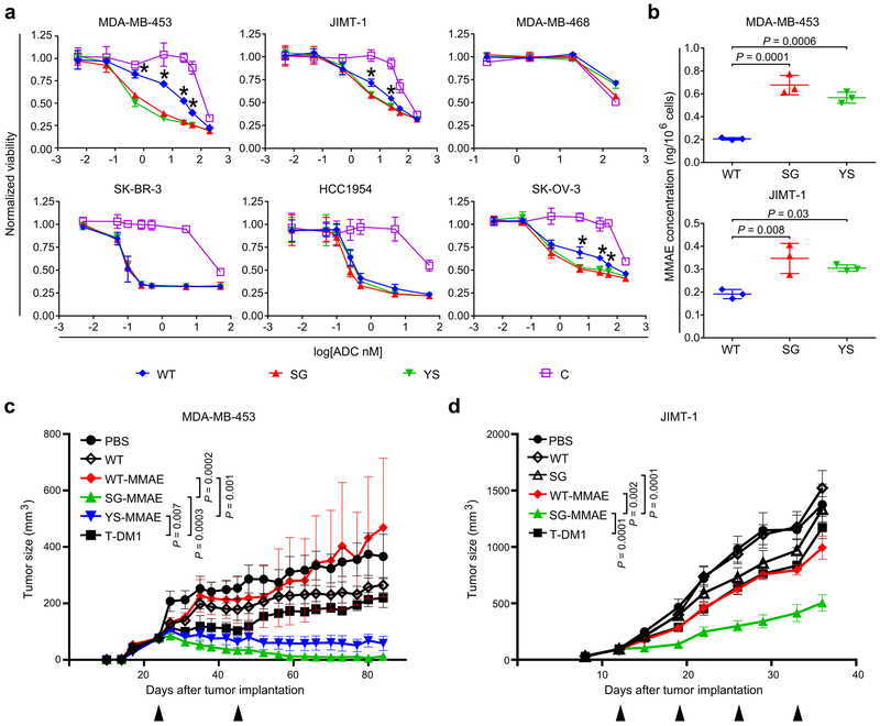
Figure 2.
ALTAs are more effective at reducing proliferation of HER2int-expressing cells and deliver increased levels of MMAE to target cells. (a) HER2-expressing cancer cells, or HER2-negative (MDA-MB-468) cells, were treated with MMAE-conjugated antibodies (WT, SG, YS or control hen egg lysozyme-specific antibody, C) for 4 days and cell viability determined. (b) MDA-MB-453 and JIMT-1 cancer cells were incubated with 10 nM MMAE-conjugated antibodies (WT, SG or YS) for 20 hours and cell-associated MMAE quantitated using LC-MS/MS. For (a) and (b), mean values of independent triplicate cell samples are shown and error bars indicate SD. Statistically significant differences between YS or SG and WT are indicated by * (one-way ANOVA with Tukey’s multiple comparison test; for panel (a), P values ranged from 0.001-0.0001 (MDA-MB-453), 0.003-0.008 (JIMT-1) and 0.001-0.05 (SK-OV-3)). (c) Female BALB/c SCID mice bearing MDA-MB-453 tumors were treated twice, with a 21 day interval (arrowheads), with 2 mg/kg ADC (WT-MMAE, SG-MMAE or YS-MMAE), T-DM1, unconjugated WT pertuzumab (WT) or vehicle (PBS) (n = 7 mice/group for YS-MMAE, T-DM1 or PBS; 8 mice/group for WT-MMAE, SG-MMAE or WT). (d) Female BALB/c SCID mice bearing JIMT-1 tumors were treated weekly (four times; arrowheads) with 2 mg/kg ADC (WT-MMAE or SG-MMAE), T-DM1, unconjugated WT or SG, or vehicle (PBS) (n = 6 mice/group for PBS; 7 mice/group for WT-MMAE, SG-MMAE, T-DM1, WT or SG). For (c) and (d), the mean tumor size for each treatment group is shown and error bars indicate SE. Statistically significant differences at the experimental endpoints are indicated by * (two-tailed Mann-Whitney U test and unpaired two-tailed t-test, respectively). For panels (a)-(d), at least two independent experiments were carried out with similar results.

