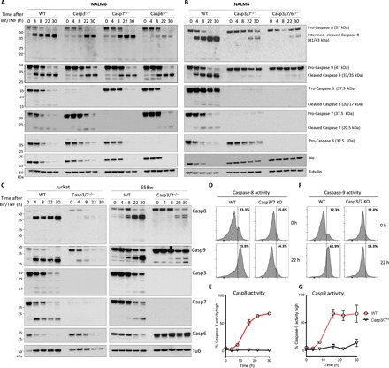
Fig. 4. Simultaneous absence of caspase-3 and -7 results in a significant decrease in caspase-8 and -9 activation in extrinsic apoptosis.

(A to C) Western blot of NALM6 wild-type (WT) and caspase-3, -7, and -6−/− cells as well as caspase-3/7−/− or caspase-3/7/6−/− cells untreated or treated with Birinapant (50 nM) and hTNF (10 ng/ml) for the indicated times. Cells were lysed and processed for Western blot. (A) Disrupting a single effector caspase-3, -7, or -6 results in no difference in caspase-8 or -9 activation. Incubation with caspase-8 antibody shows appearance of intermediate cleaved caspase-8 (41/43 kDa) and active caspase-8 (18 kDa) in WT cells and in caspase-3, -7, or -6−/−. Incubation with caspase-9 antibody shows appearance of cleaved caspase-9 (37/35 kDa) in WT cells and in caspase-3, -7, or -6−/−. Line graphs show the mean ± SEM for two repeated experiments performed in duplicate. (B) Disruption of caspase-3 and -7 simultaneously results in significant delay of the appearance of intermediate cleaved caspase-8 and no appearance of active caspase-8 as well as significant reduction and delay of cleaved caspase-9. Similar results in caspase-3/7/6−/−. (C) Jurkat WT and caspase-3/7−/− cells as well as 658w WT and caspase-3/7−/− cells show analog results. (D to G) NALM6 WT or caspase-3/7−/− cells were left untreated or treated for 22 hours with Birinapant (50 nM) and hTNF (10 ng/ml). Treated WT NALM6 cells show increased cleavage of caspase-8 (E) and caspase-9 (G) substrates over indicated time, whereas caspase-3/7−/− show no increased activity for caspase-8 and -9 substrates. Quantitation of caspase-8 and -9 activity is given (E and G), respectively. Histograms show representative data from at least two experiments performed in duplicate, with the mean from these ± SEM presented in the line graphs.
