
Fig. 2. Depletion of Zic5 renders the cells more resistant to low glucose–induced apoptosis.
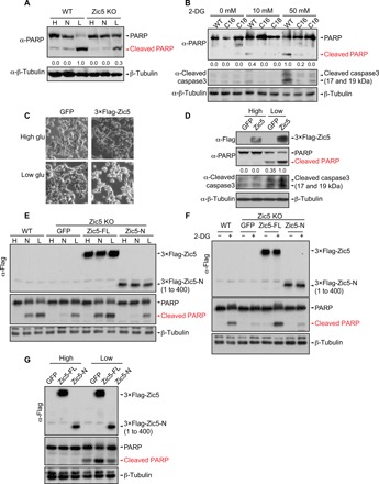
(A) Western blot showing the cleaved PARP levels of Zic5 WT and KO cells cultured in the Dulbecco’s modified Eagle’s medium (DMEM) medium with high (H, 25 mM), normal (N, 5.5 mM), or low glucose (L, 0.5 mM). (B) Western blot showing the cleaved PARP and cleaved caspase3 levels in Zic5 WT and KO cells treated with an increasing concentration of 2-DG. (C) Bright-field cellular morphology and (D) Western blot showing the cleaved PARP and cleaved caspase3 levels of HCT116 cells expressing GFP or 3×Flag-Zic5 cultured in the DMEM medium with high or low glucose. (E) Western Blot showing the cleaved PARP levels of Zic5 WT and KO cells and KO cells restored with Zic5-FL or Zic5-N cultured in the DMEM medium with high (H, 25 mM), normal (N, 5.5 mM), or low glucose (L, 0.5 mM). (F) Western blot showing the cleaved PARP levels in Zic5 WT and KO cells, and KO cells restored with Zic5-FL or Zic5-N treated with or without 50 mM 2-DG. (G) Western blot showing the cleaved PARP levels of HCT116 cells expressing green fluorescent protein (GFP), 3×Flag-Zic5-FL, or 3×-Flag-Zic5-N cultured in the DMEM medium with high or low glucose. Zic5-FL and Zic5-N were detected by antibody against Flag. Cleaved PARP signals were quantified with ImageJ.