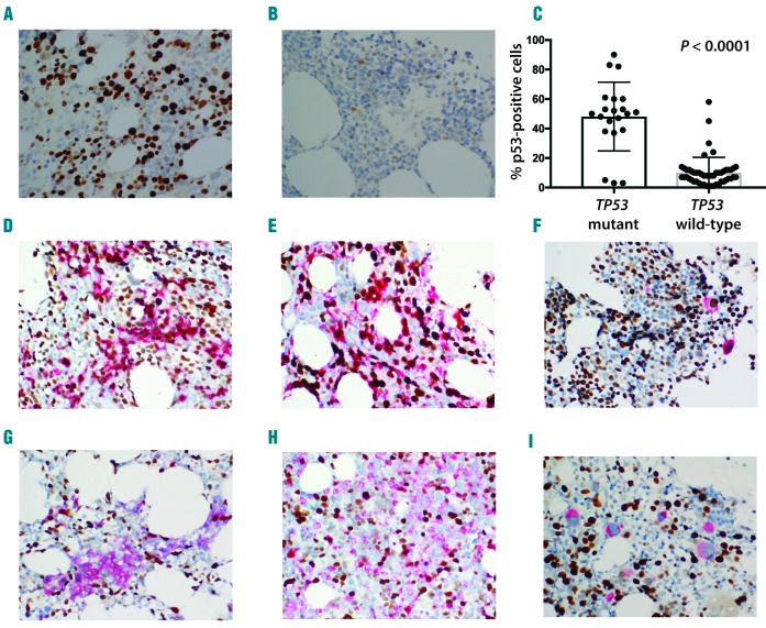
Figure 1.
TP53 expression is increased in TP53-mutated cases but does not consistently associate with specific bone marrow (BM) lineage. Representative micrographs of TP53 staining of TP53-mutated (A) and TP53 wild-type case (B). Significantly higher TP53 staining (% positive cells) in mutated versus wild-type TP53 cases (C). Co-expression of TP53 in different BM lineages (brown nuclear staining: TP53; red staining: lineage marker). High (D) and low (G) co-expression of TP53 in E-cadherin-positive erythroid elements. High (E) and low (H) co-expression of TP53 in myeloperoxidase-positive myeloid elements. High (F) and low (I) co-expression of TP53 in CD61-positive megakaryocytes.

