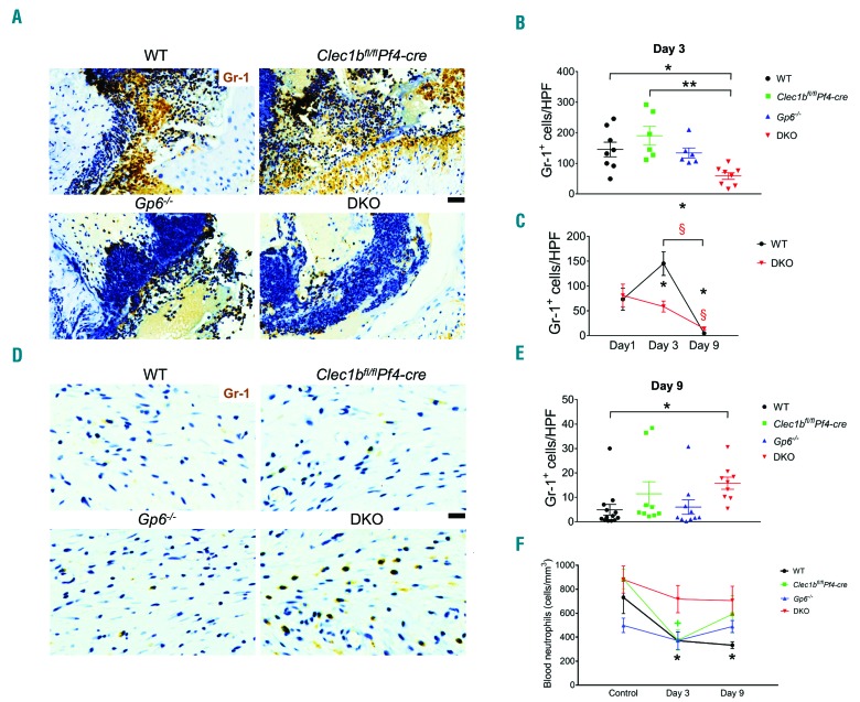
Figure 5.
Neutrophil influx is decreased during the inflammatory phase of wound healing following platelet CLEC-2 and GPVI double-deletion. (A) Detection of neutrophils (Gr-1 staining; brown) in wound at day 3 post injury. (B) Quantification of neutrophils (Gr-1+ cells) in wound at day 3 post injury. *P<0.05; **P<0.01. (C) Comparison of Gr1+ cells between day 1, day 3, and day 9 post injury in wild-type (WT) and DKO mice. P<0.05 in *WT and §DKO mice, compared to the data at day 1 post injury, respectively. The bracket shows P<0.05 for the comparison between day 3 and day 9 post injury in *WT and §DKO mice, respectively. (D) Detection of neutrophils (Gr-1 staining; brown) in wound at day 9 post injury. (E) Quantification of neutrophils (Gr-1+ cells) in wound at day 9 post injury. *P<0.05. (F) Comparison of blood neutrophil counts between baseline, day 3, and day 9 post injury in each mouse strain. P<0.05 in *WT and +Clec1bfl/flPf4-cre mice, compared to their control, respectively. Sample numbers in unchallenged control = 10, day 1 = 5, day 3 = 6-9, and day 9 post injury = 9-13, respectively. Graphs are presented as mean±Standard Error of Mean and analyzed by one-way ANOVA with Bonferroni’s multiple comparison test. Scale bar = 20 μm.

