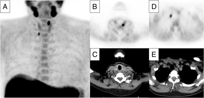
Fig. 1.
Example of a positive 18F-fluorocholine PET/CT representing a double parathyroid adenoma. Serum calcium level in this patient was 2.61 mmol/L with a parathyroid hormone level of 27.7 pmol/L. The maximum intensity projection image (a) shows two foci of intense choline uptake in the neck and physiological uptake in the salivary glands, thyroid gland, liver, spleen, and bone marrow. Axial views of PET and CT show foci suspicious for parathyroid adenomas located dorsocranial to the left thyroid lobe (b, c, dimensions 7 × 5 mm and SUVmax of 8.7) and inferior to the right thyroid lobe (d, e, dimensions 6 × 4 mm and SUVmax of 5.6)

