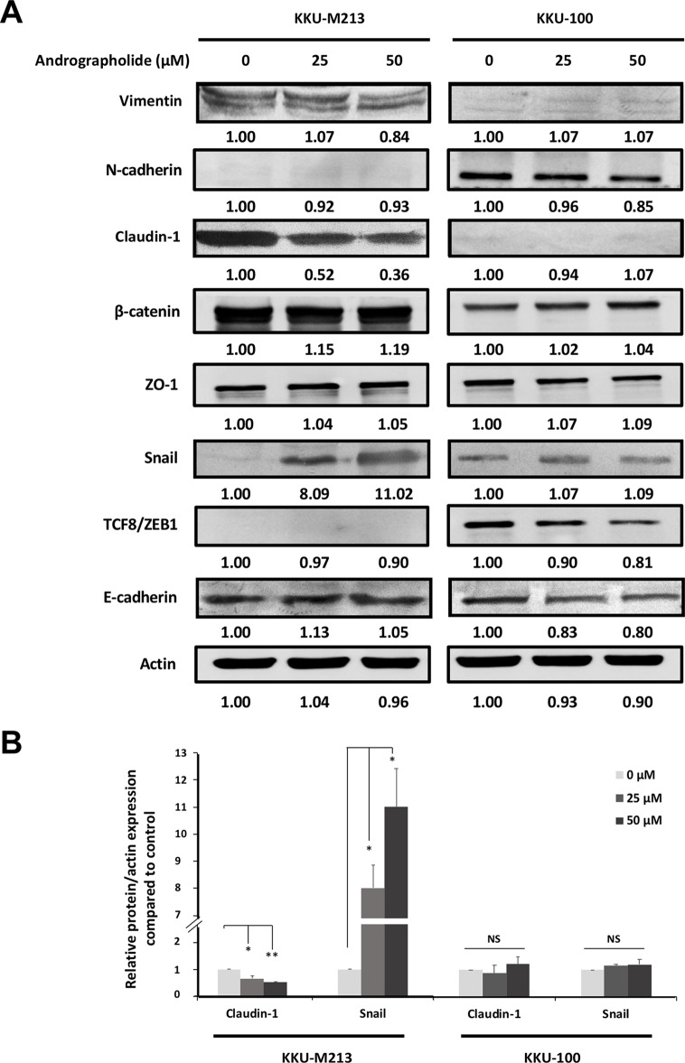
Figure 2.
Effects of andrographolide on the expression of epithelial–mesenchymal transition (EMT) proteins in CCA cells. (A) KKU-M213 and KKU-100 cells were treated with 0, 25, and 50 µM of andrographolide for 24 h prior to harvesting proteins. The expression of EMT-related proteins was evaluated using Western immunoblotting with actin as internal control. Representative results of three independent experiments are shown. The level of protein expression was quantified by intensitrometric analysis relative to actin as ratios to the untreated control. (B) The graphs present the expression of claudin-1 and Snail in KKU-M213 and KKU-100 upon the treatment of andrographolide. The results of bar graph are represented by the average of relative intensity compared with that of the control. Data were presented as mean ± SE, which were derived from three independent experiments, *p < 0.05, **p < 0.01.

