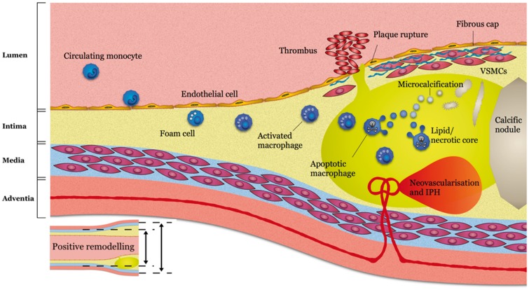
Figure 1.
Imaging targets of high-risk plaque. Reused from Adamson et al.2 Circulating monocytes migrate into early intimal thickening where they phagocytose lipid becoming foam cells and activated macrophages detectable on 68Ga-DOTATATE positron emission tomography. Vascular remodelling can be detected on computed tomography coronary angiography prior to luminal stenosis developing. As the lipid core develops this can be detected as low-density signal on computed tomography coronary angiography. The resulting hypoxic environment prompts neovascularization with friable vessels prone to intraplaque haemorrhage, both of which can be detected on magnetic resonance coronary angiography. A necrotic core develops with microvesicles arising from apoptotic macrophages and vascular smooth muscle cells giving rise to microcalcifications detectable on 18F-fluoride positron emission tomography before coalescing into more stable calcific nodules detectable on computed tomography calcium scans. Rupture of the fibrous cap may result in intraluminal thrombosis detectable on magnetic resonance coronary angiography.

