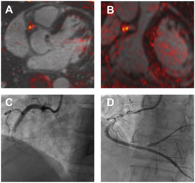
Figure 4.
Coronary atherosclerosis T1-weighted characterization with integrated anatomical reference (CATCH). T1-weighted magnetic resonance coronary angiogram of a patient who presented with an inferior myocardial infarction shows evidence of a focal high intensity lesion (arrows) in the right coronary artery on magnetic resonance imaging (A and B). Subsequent coronary angiogram demonstrated occlusion of the mid-right coronary artery (C) with restoration of flow following thrombus aspiration (D).

