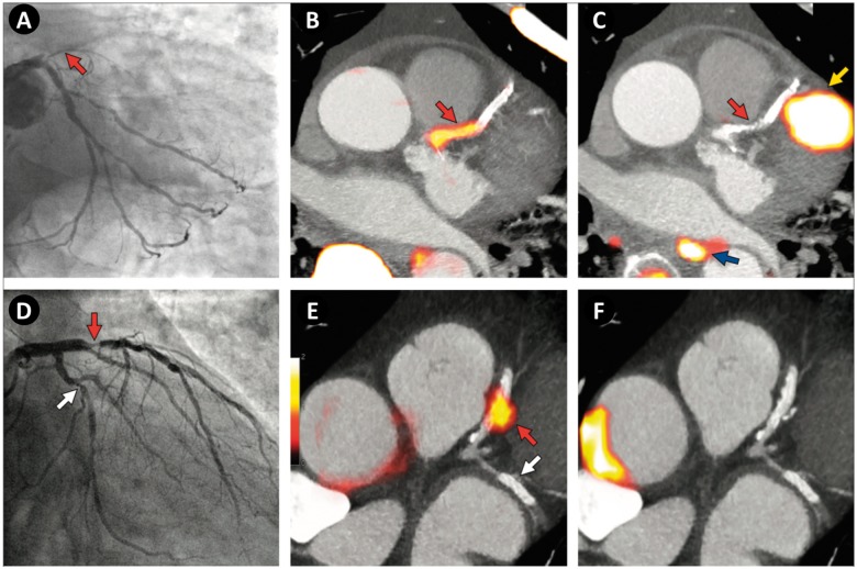
Figure 5.
Focal 18F-fluoride and 18F-fluorodeoxyglucose uptake in patients with myocardial infarction and stable angina. (Top row, A–C) Patient with acute ST-segment elevation myocardial infarction with (A) proximal occlusion (red arrow) of the left anterior descending artery on invasive coronary angiography and (B) intense focal 18F-fluoride uptake (yellow-red) at the site of the culprit plaque (red arrow) on the combined positron emission and computed tomography coronary angiography (PET-CTCA). Corresponding 18F-fluorodeoxyglucose PET-CT image (C) showing no uptake at the site of the culprit plaque. Note the significant myocardial uptake overlapping with the coronary artery (yellow arrow) and uptake within the oesophagus (blue arrow). (Bottom row) Patient with anterior non-ST-segment elevation myocardial infarction with (D) culprit (red arrow; left anterior descending artery) and bystander non-culprit (white arrow; circumflex artery) lesions on invasive coronary angiography that were both stented during the index admission. Only the culprit lesion had increased 18F-NaF uptake on PET-CT (E) after percutaneous coronary intervention. Corresponding 18F-fluorodeoxyglucose PET-CT (F) showing no uptake either at the culprit or the bystander stented lesion. Note intense uptake within the ascending aorta. Adapted from Joshi et al.47

