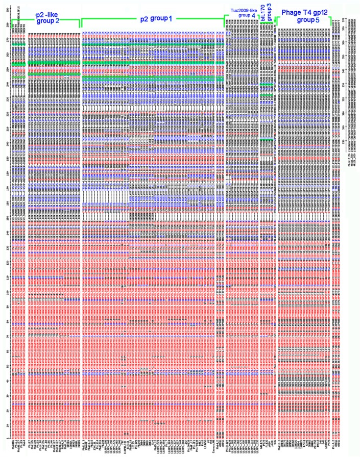
Figure 2.
Sequence alignment of the RBP proteins. The various RBP groups, based on the predicted structure of the RBP head domain, are identified on the right of the alignment. The conserved amino acids are in red and the partially conserved in blue. Otherwise in black. The phage p2 4 essential amino-acids for receptor binding, Trp144, His232, Asp234 and Arg256, are conserved in the p2 class and only partly in the p2-like class. They are highlighted in green. This is not a cited image. Multalin is a program Performed with Multalin.

