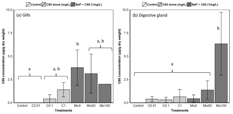
Figure 2.
Liquid chromatography-mass spectrometry (LC–MS) analyses of C60 in M. galloprovincialis (a) gills and (b) digestive gland (means ± SE). Data marked with different letters differed significantly (Tukey post-hoc test; p < 0.05). An analytical problem led to the loss of two samples of the gills from mussels exposed to Mix100 explaining the absence of standard error.

