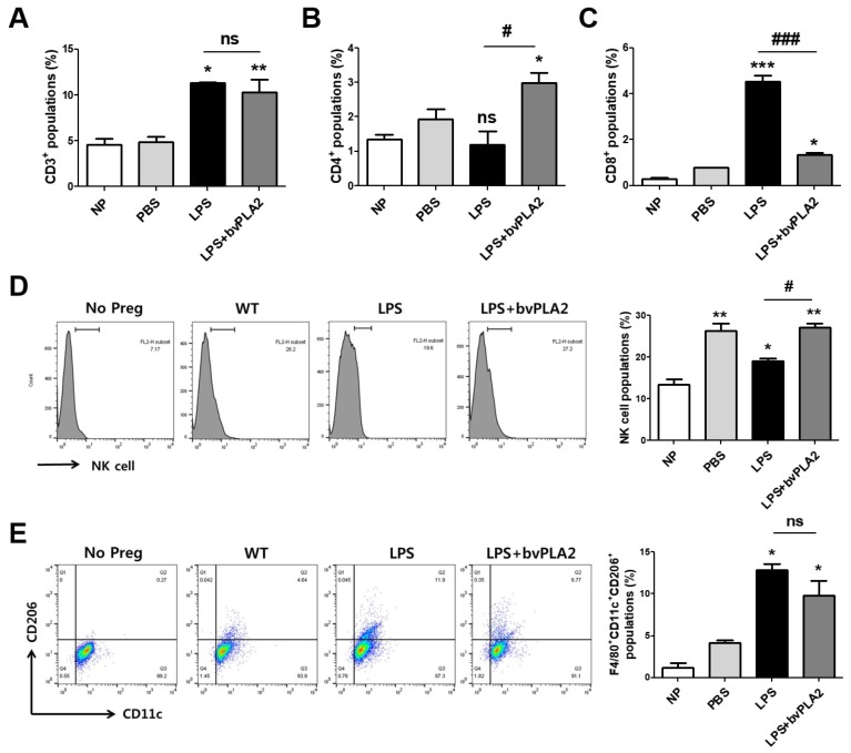
Figure 4.
Modulation of immune cell types in LPS-induced abortion mice exposed to bvPLA2. Monocytes were isolated from the uterine tissues of female mice on GD 11.5 after treatment with PBS, LPS, and LPS + bvPLA2 or from non-pregnant females. The percentages of (A) CD3+ total T cells, (B) CD4+ T helper cells, and (C) CD8+ cytotoxic T cells from the uterine tissues are depicted. (D) Representative histogram and the percentage of DBA-lectin+ uNK cells stained and analyzed by flow cytometry. (E) Dot plot and the percentage of F4/80+CD11c+CD206+ M2 macrophages stained and analyzed by flow cytometry. Significance: * p < 0.05, ** p < 0.01, and *** p < 0.001 vs. the NP group; # p < 0.05 and ### p < 0.001 vs. the LPS group; ns: no significance.

