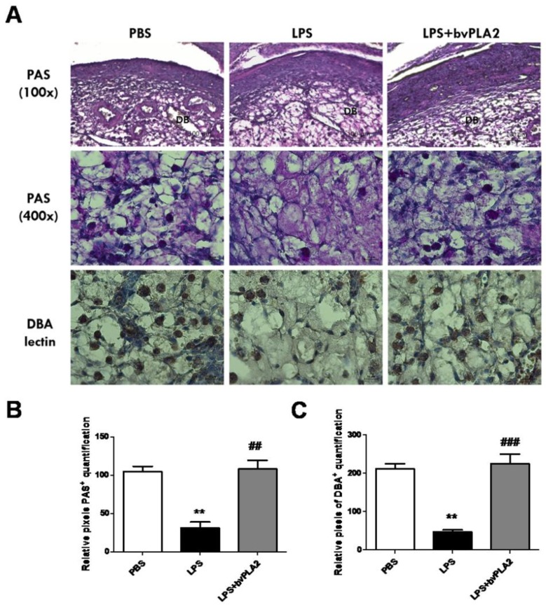
Figure 5.
Alteration of uterine natural killer (uNK) cells in the mouse uterus exposed to bvPLA2. (A) uNK cells were determined by periodic acid–Schiff (PAS) staining (100× upper and 400× lower) and immunohistochemical staining with biotinylated Dolichos biflorus agglutinin (DBA) lectin in the uterine tissues. Scale Bars: 100 μm for PAS (100×) panel and 20 μm for PAS (400×) panel. The figure represents sections from five individual mice. DB: decidua basalis. Bar graphs of data pertaining to the effects of bvPLA2 pretreatment on uNK cells by (B) PAS staining and (C) DBA lectin. Significance: ** p < 0.01 vs. the PBS group; ## p < 0.01 and ### p < 0.001 vs. the LPS group.

