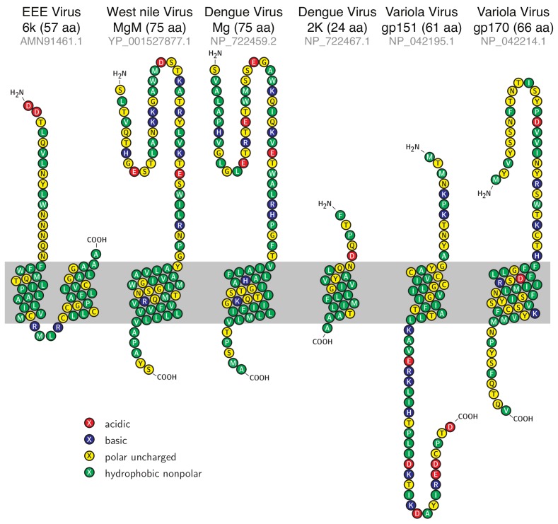
Figure 1.
Sequence and topology of candidate viroporins. Sequences of the proteins analyzed in the current study alongside their predicted topologies according to Phobius [39,40]. The shaded region indicates the presumed position of the lipid bilayer. The lengths of the proteins are stated in the parentheses and the accession numbers in gray. The figure was prepared using TEXtopo version 1.5 [56].

