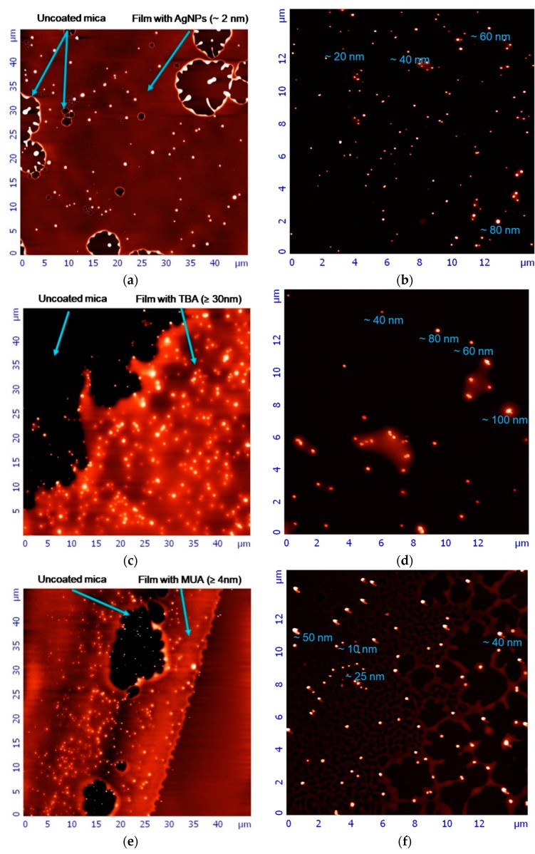
Figure 2.
Atomic force microscopy (AFM) images of newly synthesized AgNPs. (a) Images of naked AgNPs precipitated on a mica after 100-fold dilution. Arrows indicate the uncoated mica and the polymer layer height; (b) images of naked AgNPs precipitated on the uncoated mica after 100-fold dilution; (c) images of AgNP-TBA nanoparticles precipitated on a mica from the original solution. Arrows indicate the uncoated mica and the polymer layer height; (d) images of AgNP-TBA nanoparticles precipitated on the uncoated mica from the original solution; (e) images of AgNP-MUA nanoparticles precipitated on a mica after 30-fold dilution. Arrows indicate the uncoated mica and the polymer layer height; (f) images of AgNP-MUA nanoparticles precipitated on the uncoated mica after 30-fold dilution. Nanoparticles’ heights are given below measured individuals.

