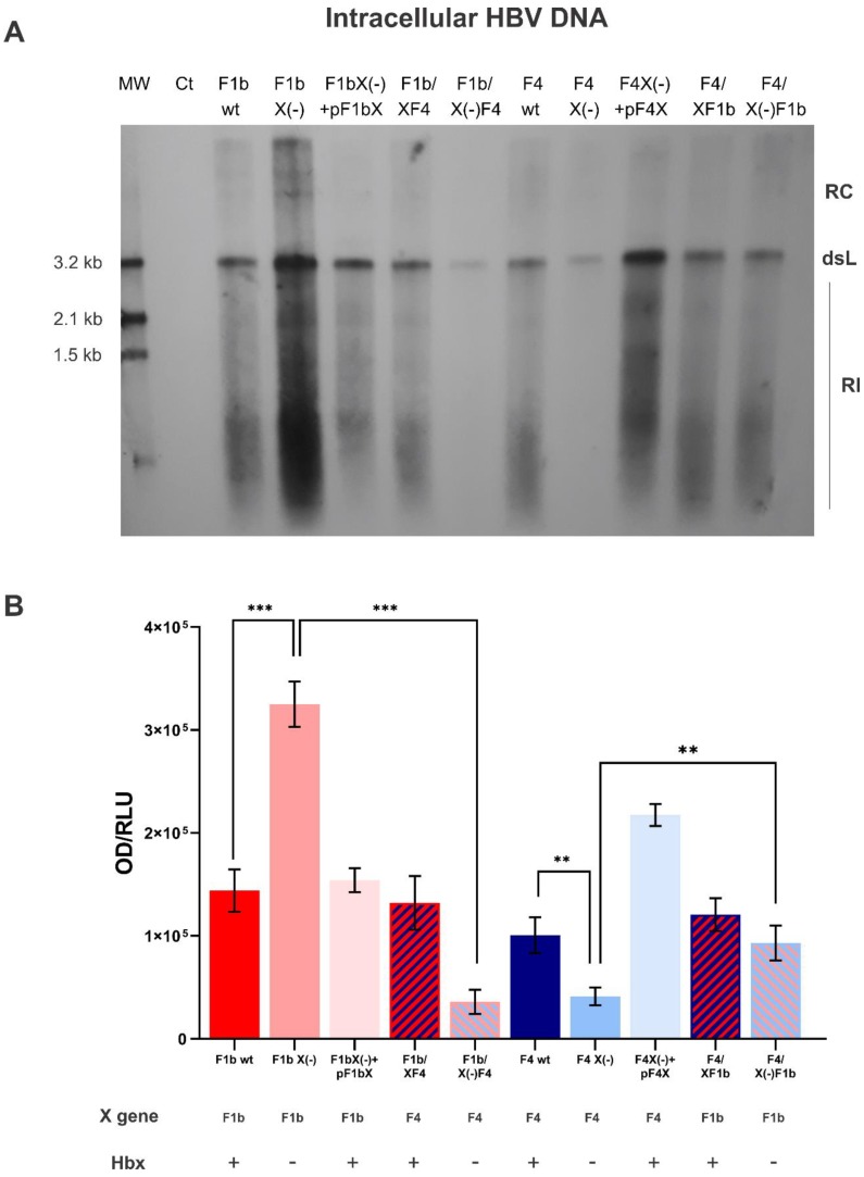
Figure 2.
Analysis of intracellular HBV DNA of sgtF1b and sgtF4 variants. HuH-7 cells were transfected with linear full-length HBV genomes of pCH-9/3091 POL minus plasmid (Ct), F1b wt, F1b X(-), F1b X(-)+pF1bX, F1b/XF4, F1b/X(-)F4, F4 wt, F4 X(-), F4 X(-)+pF4X, F4/XF1b and F4/X(-)F1b variants. Ninety-six hours post-transfection, cells were harvest and the amount of HBV DNA replicative intermediates was assessed by Southern blot analysis (A). The DNA input control (Ct) was not visible, which shows no input interference in the Southern blot analysis. RC: HBV relaxed circular DNA; dsL: HBV double-stranded linear DNA; RI: HBV replicative intermediates. (B) Relative intensity of the bands was quantified using ImageJ software. Results were expressed in relation to luciferase relative light units (RLU). Shown values represent the mean ± standard deviation of three independent experiments. ** p < 0.005 and *** p < 0.0001.

